Anatomy Of Musckes Sndctendons - Anatomy Physiology Illustration. The muscles of the abdomen, lower back, and pelvis are separated from those of the chest by the muscular wall of the diaphragm, the critical breathing muscle. This important tendon in the back of the calf and ankle stores the elastic. Tendon, tissue that attaches a muscle to other body parts, usually bones. The hands enable us to perform many of our daily activities such as driving, writing and cooking. By contracting, they also aid the venous return of blood to the heart and with age, these components of the musculoskeletal system progressively degenerate, which contributes to frailty and increases the risk of falls and fractures.
As these muscles contract and relax, they move skeletal bones to create movement of the body. The right scapula from the front and back side. The smaller bone that runs alongside the tibia (fibula) and the kneecap (patella) are the other bones that make the knee joint. The quad muscles— which form the meaty mass on the front of your thighs — are among your strongest muscle groups, and play a critical role in athletic activities. By connecting our rigid bones to our powerful muscles, tendons allow us to move.
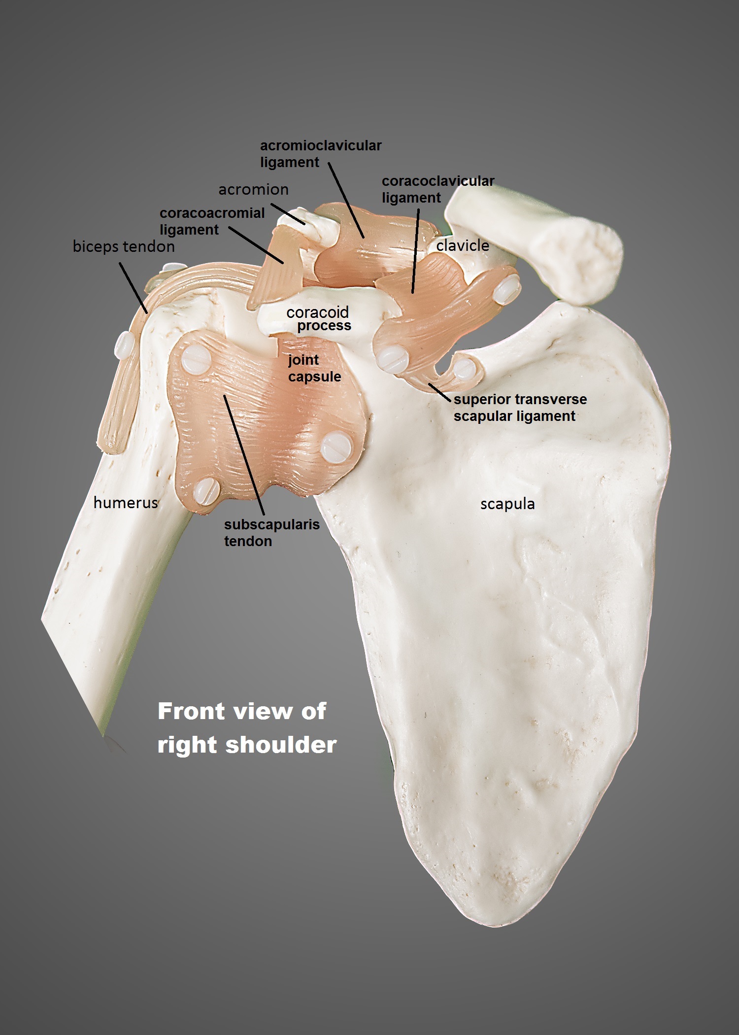
Anatomy Of The Shoulder Ut Health San Antonio from www.uthscsa.edu The primary function of the shoulder girdle is to give strength and range of motion to the arm. The shoulder is not a single joint, but a complex arrangement of bones, ligaments, muscles, and tendons that is better called the shoulder girdle. By contracting, they also aid the venous return of blood to the heart and with age, these components of the musculoskeletal system progressively degenerate, which contributes to frailty and increases the risk of falls and fractures. Related posts of muscles and tendons of the leg muscle anatomy forearm. Extensor carpi radialis brevis extensor carpi radialis longus Insertion for gluteus maximus and tensor fascia latae connective. The right scapula from the front and back side. The human hand is made up of the wrist, palm, and fingers and consists of 27 bones, 27 joints, 34 muscles, over 100 ligaments and tendons, and many blood vessels and nerves.
Tendon, tissue that attaches a muscle to other body parts, usually bones.
The primary function of the shoulder girdle is to give strength and range of motion to the arm. The tightening and relaxing of the calf muscles enables the ankle to bend downward and upward. On the other hand, the insertion is where a tendon attaches that muscle to the *more* movable bone. The muscles of the abdomen, lower back, and pelvis are separated from those of the chest by the muscular wall of the diaphragm, the critical breathing muscle. All together they help hold your upper arm in place in the shoulder. By connecting our rigid bones to our powerful muscles, tendons allow us to move. Find the best weight lifting exercises that target each muscle or groups of muscles. There are three layers of gluteal muscles on the posterior hips, just like there are three layers of muscles in the abdominal trunk. The tendon is firmly connected to muscle fibres at one end and to components of the bone at its other end. The human hand is made up of the wrist, palm, and fingers and consists of 27 bones, 27 joints, 34 muscles, over 100 ligaments and tendons, and many blood vessels and nerves. This important tendon in the back of the calf and ankle stores the elastic. The fleshy, thick part of the muscle is called its belly. Tendons connect the knee bones to the leg muscles that move the knee.
All together they help hold your upper arm in place in the shoulder. It forms the floor of the cubital fossa. The tendon is firmly connected to muscle fibres at one end and to components of the bone at its other end. The red lines show where the tendons attach the muscles to the bones. The calf muscles (gastrocnemius and soleus), which are connected to the calcaneus via the achilles tendon.
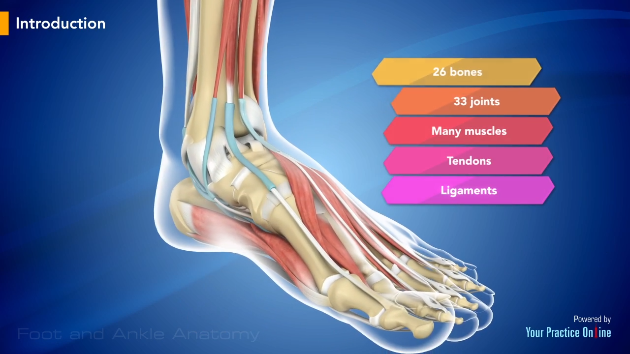
Foot And Ankle Anatomy Video Medical Video Library from www.ypo.education Skeletal muscles are attached to bones by tendons and can be as long as 30 cm, although they are usually 2 to 3 cm in length. The shoulder is not a single joint, but a complex arrangement of bones, ligaments, muscles, and tendons that is better called the shoulder girdle. In this lesson, we look at the muscle. Muscles and tendons of upper leg. By connecting our rigid bones to our powerful muscles, tendons allow us to move. Every skeletal muscle has three main parts: The majority of muscles in the leg are considered long muscles, in that they stretch great distances. Insertion for gluteus maximus and tensor fascia latae connective.
The quad muscles— which form the meaty mass on the front of your thighs — are among your strongest muscle groups, and play a critical role in athletic activities.
Insertion for gluteus maximus and tensor fascia latae connective. Tendon, tissue that attaches a muscle to other body parts, usually bones. The quadriceps muscles provide strength and power with knee extension (straightening). Four muscles and their attached tendons make up the rotator cuff. Tendons attach muscle to bone. By contracting, they also aid the venous return of blood to the heart and with age, these components of the musculoskeletal system progressively degenerate, which contributes to frailty and increases the risk of falls and fractures. Tendons are cords made of tough tissue, and they work as special connector pieces between bone and muscle. The shoulder is not a single joint, but a complex arrangement of bones, ligaments, muscles, and tendons that is better called the shoulder girdle. The hands enable us to perform many of our daily activities such as driving, writing and cooking. Together, these muscles straighten your knee, stabilize your knee joint, assist in flexing your hip (drawing your knee towards your chest), and help absorb force when you land after jumping or leaping. Ligaments connect two or more bones together and help stabilize joints. The quad muscles— which form the meaty mass on the front of your thighs — are among your strongest muscle groups, and play a critical role in athletic activities. *the origin, insertion, and belly.* a muscle's origin is where a tendon attaches it to the *less* movable bone.
Tendons vary in size and are somewhat elastic and attach bones to muscles. Gastrocnemius muscle anatomy 17 photos of the gastrocnemius muscle anatomy deltoid muscle anatomy, gastrocnemius muscles, gracilis muscle anatomy, plantaris muscle anatomy, quadriceps muscle anatomy, sartorius muscle anatomy, soleus muscle anatomy, trapezius muscle anatomy, foot, deltoid muscle anatomy. Muscles and tendons of upper leg. Tendons are the connective tissues that transmit the mechanical force of muscle contraction to the bones; The image below shows the bones of the hand from the back side.
Muscle Anatomy Of The Plantar Foot Everything You Need To Know Dr Nabil Ebraheim Youtube from i.ytimg.com Movement occurs when our muscles pull on our bones, relocating them. The quadriceps muscles provide strength and power with knee extension (straightening). By contracting, they also aid the venous return of blood to the heart and with age, these components of the musculoskeletal system progressively degenerate, which contributes to frailty and increases the risk of falls and fractures. Tendons connect the knee bones to the leg muscles that move the knee. The tendon is firmly connected to muscle fibres at one end and to components of the bone at its other end. The muscles of the abdomen, lower back, and pelvis are separated from those of the chest by the muscular wall of the diaphragm, the critical breathing muscle. The primary function of the shoulder girdle is to give strength and range of motion to the arm. Tendons are the connective tissues that transmit the mechanical force of muscle contraction to the bones;
Tendons are cords made of tough tissue, and they work as special connector pieces between bone and muscle.
Lesson on the anatomy of the forearm: The quad muscles— which form the meaty mass on the front of your thighs — are among your strongest muscle groups, and play a critical role in athletic activities. The posterior tibialis muscle, which supports the arch of the foot and enables the foot to turn. The shoulder is not a single joint, but a complex arrangement of bones, ligaments, muscles, and tendons that is better called the shoulder girdle. Four muscles and their attached tendons make up the rotator cuff. The right scapula from the front and back side. Movement occurs when our muscles pull on our bones, relocating them. Find the best weight lifting exercises that target each muscle or groups of muscles. Tendons are thick bands of tissue that connect muscles to bones. The tightening and relaxing of the calf muscles enables the ankle to bend downward and upward. The hands enable us to perform many of our daily activities such as driving, writing and cooking. Most of the muscles which act on the wrist joint are situated within the forearm, with only the tendon crossing the joint and inserting on the hand. Lesson on the anatomy of the forearm:
Share :
Post a Comment
for "Anatomy Of Musckes Sndctendons - Anatomy Physiology Illustration"


Post a Comment for "Anatomy Of Musckes Sndctendons - Anatomy Physiology Illustration"